44 basic animal cell diagram with labels
sciencequiz.net › newjcscience › jcbiologyThe Cell - ScienceQuiz.net One of the differences between a plant cell and an animal cell is that plant cells have a cell wall while animal cells do not. animal cells contain chloroplasts while plant cells do not. Structure of Cell: Definition, Types, Diagram, Functions - Embibe Exams It is spherical in animal cells and is located at the centre. The nucleus has a nuclear envelope, nuclear sap, nuclear matrix, chromatin and nucleolus. The nuclear envelope separates the nucleus from the cytoplasm, it has many pores (the nuclear pores) and encloses the liquid ground substance, the nucleoplasm.
Eukaryotic Cell: Definition, structure and organelles - Kenhub The eukaryotic cells types are generally found in animals, plants, algae, and fungi. For the purpose of this article, the primary focus will be the structure and histology of the animal cell. The major differences between animal and plant cells will be explored as well. As previously stated, the fundamental components of a cell are its organelles.

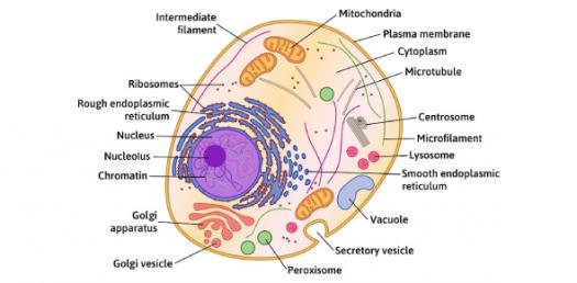
Basic animal cell diagram with labels
[Plant Cell Without Labels] - 15 images - game statistics plant cell ... [Plant Cell Without Labels] - 15 images - animal and plant cells both have a cell membrane cytoplasm and a, this is the cell wall it protects the plant cell from an, blog kami biology form 4 animal cell plant cell, ideas and class info biology, Histology guide: Definition and slides | Kenhub The cells are laid down on top of dense irregular connective tissue, the basement membrane (BM). Epithelium is classified by both it's cellular morphology and the number of cell layers. Based on morphology, epithelial cells can be either squamous (flat), cuboid (cube) or columnar (rectangular). › 37006818 › Junqueiras_BasicJunqueira's Basic Histology Text and Atlas, 14th Edition Junqueira's Basic Histology Text and Atlas, 14th Edition. Marwan Othman. Download Download PDF. Full PDF Package Download Full PDF Package. This Paper. A short ...
Basic animal cell diagram with labels. 43.5B: Cleavage, the Blastula Stage, and Gastrulation The cells in the blastula rearrange themselves spatially to form three layers of cells in a process known as gastrulation. During gastrulation, the blastula folds upon itself to form the three layers of cells. Each of these layers is called a germ layer, which differentiate into different organ systems. Figure 43.5 B. 1: Cell Biology Quiz - ProProfs Quiz A cell is the basic fundamental unit of all biological organisms. This challenging biology test on cells is designed to test your knowledge on this topic to the extremes. Good Luck with this quiz! We Challenge you to score the highest marks by answering most questions on this quiz! Questions and Answers. 1. 24.1B: Fungi Cell Structure and Function - Biology LibreTexts Figure 24.1 B. 1: Division of hyphae into separate cells: Fungal hyphae may be (a) septated or (b) coenocytic (coeno- = "common"; -cytic = "cell") with many nuclei present in a single hypha. A bright field light micrograph of (c) Phialophora richardsiae shows septa that divide the hyphae. en.wikipedia.org › wiki › Venn_diagramVenn diagram - Wikipedia A Venn diagram is a widely used diagram style that shows the logical relation between sets, popularized by John Venn in the 1880s. The diagrams are used to teach elementary set theory , and to illustrate simple set relationships in probability , logic , statistics , linguistics and computer science .
21.2B: The Lytic and Lysogenic Cycles of Bacteriophages When infection of a cell by a bacteriophage results in the production of new virions, the infection is said to be productive. Figure 21.2 B. 1: Lytic versus lysogenic cycle: A temperate bacteriophage has both lytic and lysogenic cycles. In the lytic cycle, the phage replicates and lyses the host cell. In the lysogenic cycle, phage DNA is ... Plant Cell: Diagram, Types and Functions - Embibe Exams The Plant Cell is the most basic and basic unit of all plants. Plant cells are eukaryotic, which means they have a membrane-bound nucleus and organelles, just like animal cells. That's all there is to the similarity. In comparison to animal cells, plant cells have cell walls that surround the cell membrane. 22.2A: Basic Structures of Prokaryotic Cells - Biology LibreTexts Prokaryotes are unicellular organisms that lack organelles or other internal membrane-bound structures. Therefore, they do not have a nucleus, but, instead, generally have a single chromosome: a piece of circular, double-stranded DNA located in an area of the cell called the nucleoid. Most prokaryotes have a cell wall outside the plasma membrane. Eukaryotic Cell: Structure, Characteristics & Diagram - Embibe Exams The cell cycle in eukaryotes is divided into the following phases: a. Interphase- This phase includes i. \ ( {\rm {G1}}\) phase- It is the first phase hence named the first gap \ ( {\rm {G1}}\) phase). In this phase, the cell grows in size. All the organelles duplicate but DNA duplication is not involved here.
Animal cell Definition and Examples - Biology Online Dictionary An animal cell is the fundamental functional unit of life of animals.It is also the basic unit of reproduction. Animal cells were first observed in the 17th century when microscopy was invented. Robert Hooke, an English natural philosopher, was the first to describe microscopic pores, which he later called cells, albeit from samples of a plant cork. Organelle - Genome.gov Definition. …. An organelle is a subcellular structure that has one or more specific jobs to perform in the cell, much like an organ does in the body. Among the more important cell organelles are the nuclei, which store genetic information; mitochondria, which produce chemical energy; and ribosomes, which assemble proteins. NCERT Exemplar Class 8 Science Chapter 8 Cell Structure ... - Learn CBSE (a) The above diagram represents an animal ceil because cell is bounded by cell membrane. Cell wall is absent. (b) The above diagram represents an eukaryotic cell as it has a well organised nucleus and also other cell organelles in it. Question. 34 Label the parts A to E in the given figure. Answer. The figure given in question, represents a ... Skin: Cells, layers and histological features | Kenhub Stratum basale acts as the stem cell region for the epidermis. It consists of a mixture of simple cuboidal to columnar epithelium resting on a basement membrane. Compared to the cytoplasm, the nuclei of these cells are large, euchromatic, with prominent nucleoli giving a marked basophilia to this layer.
Label Worksheet Cell Prokaryotic Search: Label Prokaryotic Cell Worksheet. coli cell P E Prokaryotic vs Based on the above word definitions, label the cells in Model 1 and Model 2 as prokaryotic or eukaryotic This is an online quiz called Label the Prokaryotic cell There is a printable worksheet available for download here so you can take the quiz with pen and paper Partnerships for Reform through Investigative Science and ...
Plant and Animal Cell: Labeled Diagram, Structure, Function - Embibe Exams Plant and Animal Cell: The cell is the basic building block of life. Cells are responsible for all aspects of life. The number of cells in an organism determines its classification. Unicellular species have only one cell, but multicellular organisms contain many cells. ... Diagram of Plant and Animal Cell. Fig: Plant Cell. Fig: Animal Cell ...
Plasma Membrane (Cell Membrane) - Genome.gov Definition. …. The plasma membrane, also called the cell membrane, is the membrane found in all cells that separates the interior of the cell from the outside environment. In bacterial and plant cells, a cell wall is attached to the plasma membrane on its outside surface. The plasma membrane consists of a lipid bilayer that is semipermeable.
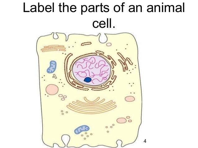
Free Printable Plant and Animal Cells Worksheets Worksheets of animal cell diagrams help your students to visually see what the animal cell looks like and identify visually the parts that make up the animal cell. Blank, Labeled, and Coloring Animal Cell Diagram - Grab these three free diagrams.

cell diagrams to label | animal cell (diagram & label)(7-2) | schooling | Pinterest | Biology ...
43.3C: Gametogenesis (Spermatogenesis and Oogenesis) Gametogenesis (Spermatogenesis and Oogenesis) Gametogenesis, the production of sperm and eggs, takes place through the process of meiosis. During meiosis, two cell divisions separate the paired chromosomes in the nucleus and then separate the chromatids that were made during an earlier stage of the cell's life cycle, resulting in gametes that ...

Questions And Answers On Labeled/Unlebled Diagrams Of A Human Cell : Label Cell Membrane Diagram ...
Cell Membrane (Plasma Membrane) - Genome.gov Definition. …. The cell membrane, also called the plasma membrane, is found in all cells and separates the interior of the cell from the outside environment. The cell membrane consists of a lipid bilayer that is semipermeable. The cell membrane regulates the transport of materials entering and exiting the cell.
Centriole - Genome.gov A centriole is a barrel-shaped organelle which lives normally within the centrosome. The centrosome is the area of the cytoplasm. It's next to the nucleus and within the centrosome. The word some refers generally to an organelle of some sort, like a lysosome or an endosome. Within that centrosome there are two centrioles.
Anatomical Terms & Meaning: Anatomy Regions, Planes, Areas, Directions Human anatomy is the study of the structure of the human body.Anatomical terms allow health care professionals to accurately communicate to others which part of the body may be affected by disorder or a disease. Terms are defined in reference to a theoretical person who is standing in what is called anatomical position (see figure below): both feet pointing forwards, arms down to the side with ...
Cell nucleus: Histology, structure and functions - Kenhub The nucleus is normally around 5-10 μm in diameter in many multicellular organisms, and the largest organelle in the cell. The smallest nuclei are approximately 1 μm in diameter and are found in yeast cells. Cell nucleus (histological slide) Mostly the shape of the nucleus is spherical or oblong.
Animal Cell: Animal Cell Diagram: Types, and Functions - Embibe Exams An animal cell is a eukaryotic cell having membrane-bound cell organelles without a cell wall. The size of the cell varies from a few microns to a few centimetres. For example, the largest animal cell is the ostrich egg measuring 170 mm x 130 mm. We can say that the size of the cell depends on the function it performs.
› mmwr › previewGuidelines for Safe Work Practices in Human and Animal ... Jan 06, 2012 · The guidelines in this section are combined biosafety best practices for both human autopsy and human surgical pathology and animal necropsy and veterinary surgical pathology. When necessary, biosafety guidelines specific for human or animal diagnostic laboratory settings are highlighted. 5.1. Autopsy/Necropsy–Associated Infections
› innovation › galvanic-cell-workGalvanic Cell: Definition, Diagram and Working - Science ABC Jan 17, 2022 · Galvanic Cell vs Electrolytic Cell. Lastly, once dead, galvanic cells cannot be revived or recharged. This is why one must change the batteries in an alarm clock or remote control from time to time. The kind of electrochemical cell that can be recharged is an electrolytic cell.
Cell Cycle - Genome.gov Cell cycle is the name we give the process through which cells replicate and make two new cells. Cell cycle has different stages called G1, S, G2, and M. G1 is the stage where the cell is preparing to divide. To do this, it then moves into the S phase where the cell copies all the DNA. So, S stands for DNA synthesis.
› proteins › proteinProtein Targeting (With Diagram) | Molecular Biology ADVERTISEMENTS: Let us make an in-depth study of the protein targeting. After reading this article you will learn about: 1. Introduction to Protein Targeting 2. Signal Sequence 3. Transport of Proteins into ER 4. Signal Sequence Recognition Mechanism 5. Role of Golgi Complex in Protein Transportation 6. Transport of Proteins from Golgi to Lysosomes 7. […]
animal cell diagram - More About Row The diagram like the one above will include labels of the major parts of an animal cell including the cell membrane nucleus ribosomes mitochondria vesicles and cytosol. Thousands of new high-quality pictures added every day. Blank Labeled and Coloring Animal Cell Diagram Grab these three free diagrams.
Diagram Respiration Label Cellular The Anatomy of a model cell part 2 learning goal Write and label equations for cellular respiration and Page 15/20 Complete this diagram Separating and labeling of pieces allow students to keep track of what molecule(s) goes into each stage, what molecule(s) are formed during each stage Cellular'Respiration'Foldable'Directions:+' 1 Cellular ...
en.wikipedia.org › Cell_membrane_(diagrammatic)Wikipedia:Featured picture candidates/Cell membrane ... Image:Plant_cell_structure_svg.svg, a Featured Picture, is under the same threat of summary deletion, as are many of LadyofHats (Mariana Ruiz) other contributions, for example Image:Human arm bones diagram.svg a FP, Image:Average prokaryote cell- en.svg, a FP, Image:Animal cell structure.svg, the FPC below...


Post a Comment for "44 basic animal cell diagram with labels"