45 dissecting microscope diagram with labels
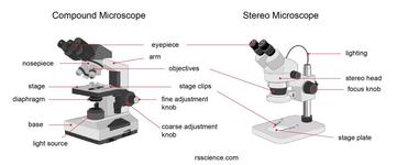
Compound Light/Dissecting Microscope Diagram | Quizlet Used to examine material mounted on microscope slides (usually thinly sectioned & stained) Provides total magnification of 40x-1000x No space for dissection Rules TRANSPORT Arm & base USE Always start at 4x, Coarse focus, Fine focus Then change objectives & use fine focus as needed Coarse focus ONLY with 4x! CLEANING Objectives/Oculars Dissecting Microscope Parts And Functions. All You Need To Know The dissecting microscope is also referred to as a stereoscopic microscope and is ordinarily used to study three-dimensional objects. And also as the name suggests for dissecting and analysing biological specimens under low magnification between two and two hundred and fifty times.
Label the microscope — Science Learning Hub Use this with the Microscope parts activity to help students identify and label the main parts of a microscope and then describe their functions. Drag and drop the text labels onto the microscope diagram. If you want to redo an answer, click on the box and the answer will go back to the top so you can move it to another box.

Dissecting microscope diagram with labels
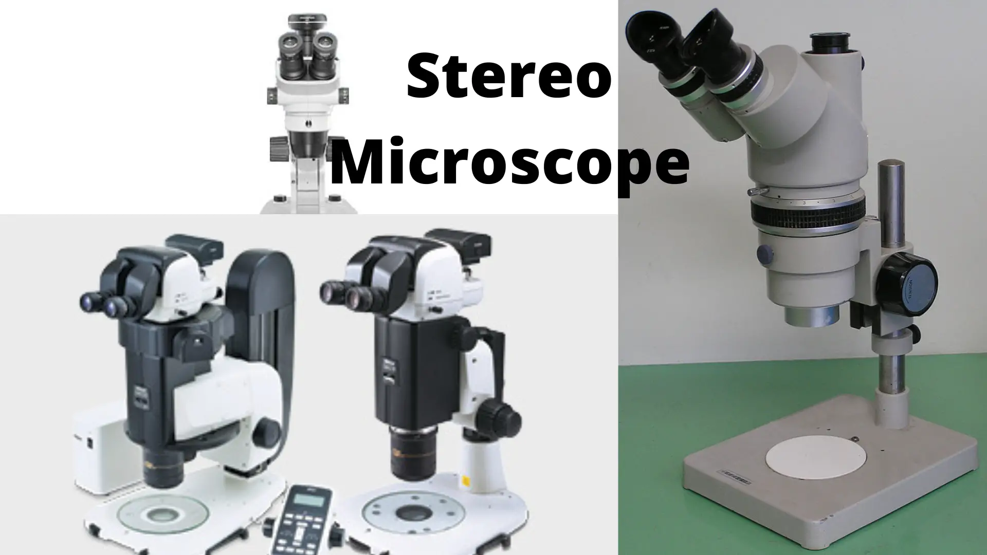
A Study of the Microscope and its Functions With a Labeled Diagram ... A Study of the Microscope and its Functions With a Labeled Diagram To better understand the structure and function of a microscope, we need to take a look at the labeled microscope diagrams of the compound and electron microscope. These diagrams clearly explain the functioning of the microscopes along with their respective parts. Dissecting microscope (Stereoscopic or Stereo microscope) This microscope is a dual-powered dissecting microscope of 10x-30x with an ability to rotate 360° making it ideal for viewing and focussing better to view samples. By rotating the lenses, users can change the magnification of image. Dissecting the treatment-naive ecosystem of human melanoma … WebJul 07, 2022 · Introduction. Melanoma brain metastases (MBMs) are the third most common cause of brain metastases after carcinomas of the lung and breast (Eichler et al., 2011) and lead to significant morbidity and mortality (Davies et al., 2011).While treatment with combination immune checkpoint blockade can be effective in patients with MBM (Tawbi …
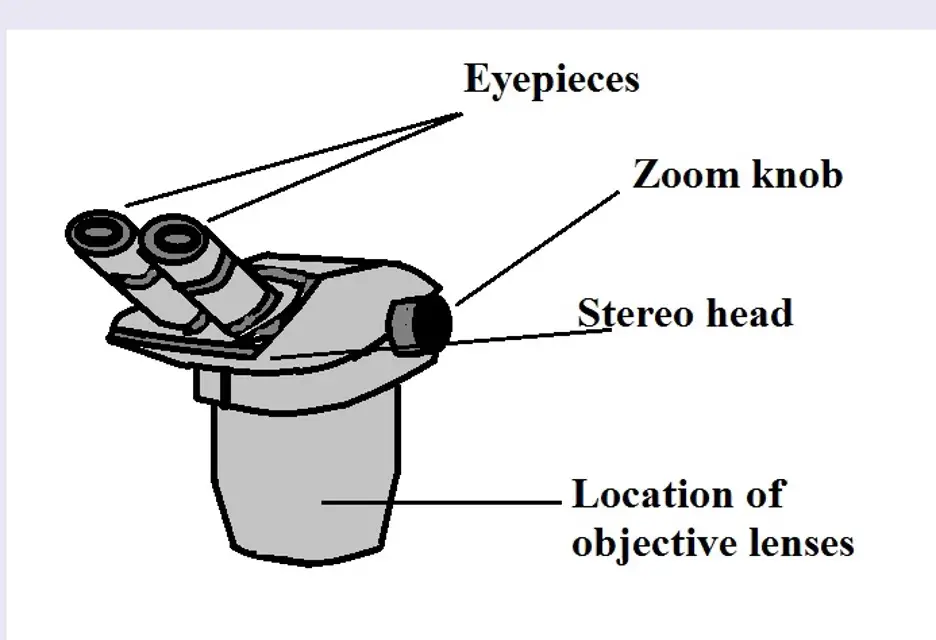
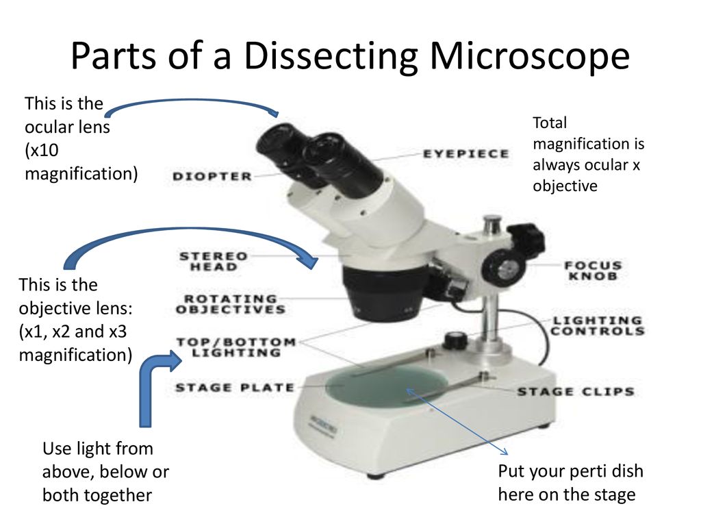
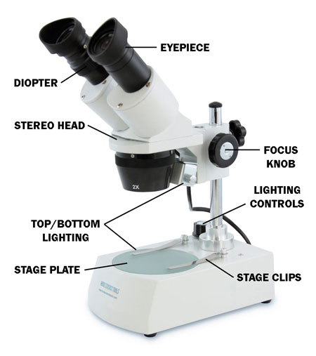
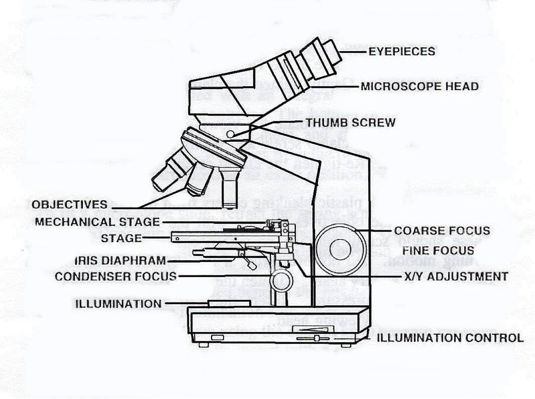
Dissecting microscope diagram with labels. Microscope Label Diagram - 34 label diagram of microscope labels ... Microscope Label Diagram - 18 images - unbiology6, biology 521 resources, label a microscope teaching resources, quia protist vocabulary, Menu ≡ ╳ Home K To 12 Science Grade 7 Learners Material - Module WebRead and do the activities in the section on How to Use The Light Microscope before performing Activity 2. Activity 2 Investigating plant cells Objectives In this activity, you should be able to: 1. prepare a wet mount; 2. describe a plant cell observed under the light microscope; 3. stain plant cells; 4. Label the microscope Diagram | Quizlet Diaphragm. Regulates the amount of light on the specimen. Light Source. Projects light upwards through the diaphragm, the specimen, and the lenses. Arm. supports the body tube. Stage. Supports the slide being viewed. Coarse Adjustment. Parts of Stereo Microscope (Dissecting microscope) – labeled diagram ... WebLabeled part diagram of a stereo microscope ... (based on color bands and their respective labels), the objectives of a dissecting microscope are located in a cylindrical cone and, therefore, are not directly seen. For the stereo microscope that comes with multiple objective lens sets (fixed power style), the cylindrical cone can be turned to ...
Low-Dose Radiotherapy Reverses Tumor Immune Desertification … WebJan 01, 2022 · Response to immune checkpoint blockade (ICB) is robust and durable in a proportion of patients. Patients with so-called cold or immune-desert tumors are, however, less likely to respond to ICB ().Important efforts are thus under way to identify effective and feasible approaches for inflaming these tumors ().Moreover, the inherent plasticity of … Dissecting microscope (Stereo or stereoscopic microscope)- Definition ... Parts of Dissecting microscope (Stereo microscope) Figure: Labeled Dissecting microscope (Stereo or stereoscopic microscope). Image created using biorender.com LED illuminators- For some of the dissecting Microscopes, they have an inbuilt LED illuminator as a source of light. Compound Microscope Parts, Functions, and Labeled Diagram Compound Microscope Definitions for Labels. Eyepiece (ocular lens) with or without Pointer: The part that is looked through at the top of the compound microscope. Eyepieces typically have a magnification between 5x & 30x. Monocular or Binocular Head: Structural support that holds & connects the eyepieces to the objective lenses. Microscope Types (with labeled diagrams) and Functions These microscopes work on the principle called contrast-enhancing technique that is utilized to produce high-contrast images to view them with more accuracy and clarity. Phase-contrast microscope labeled diagram Phase-contrast microscope functions: Its applications areas include In cases where the specimen is colorless and is very tiny
Parts of Dissecting Microscope | Botany - Biology Discussion Dissecting microscope is used to dissect small organisms or organs, e.g., embryo dissection. Its special utility is to observe such materials where high magnification is not needed. Design of Compound Microscope (With Diagram) | Biology Labelled Diagram of Compound Microscope › cell › fulltextDissecting the treatment-naive ecosystem of human melanoma ... Jul 07, 2022 · Here, we built a multi-omic single-cell atlas of treatment-naive MBMs and extracranial melanoma metastases (ECMs). Integrated analyses of this atlas coupled with validation in pre-clinical models and additional patient cohorts identified genomic, adaptive, and tumor-microenvironmental features enriched in MBM ecosystems. rsscience.com › stereo-microscopeParts of Stereo Microscope (Dissecting microscope) – labeled ... Stereo microscopes (also called Dissecting microscope) are branched out from other light microscopes for the application of viewing "3D" objects. These include substantial specimens, such as insects, feathers, leaves, rocks, sand grains, gems, coins, and stamps, etc. Functionally, a stereo microscope is like a powerful magnifying glass. Microscope labeled diagram - SlideShare 1. The Microscope Image courtesy of: Microscopehelp.com Basic rules to using the microscope 1. You should always carry a microscope with two hands, one on the arm and the other under the base. 2. You should always start on the lowest power objective lens and should always leave the microscope on the low power lens when you finish using it. 3.
Make: Science Room - Choosing a microscope - Make:
› createJoin LiveJournal Password requirements: 6 to 30 characters long; ASCII characters only (characters found on a standard US keyboard); must contain at least 4 different symbols;

Stereo Microscope: Uses, Advantages, and Disadvantages ...
EOF

Simple Microscope - Diagram (Parts labelled), Principle ...
COVID-19 immune features revealed by a large-scale single-cell ... WebApr 01, 2021 · IGHV genes differentially used by moderate or severe COVID-19 patients compared with healthy controls and their intersections are shown with different colors. Venn diagram is used to show their overlaps with those published SARS-CoV-2 antibodies. Adjusted p values < 0.05 are indicated (two-sided unpaired Wilcoxon test).

labeling dissecting microscope Diagram | Quizlet
journals.plos.org › plosgenetics › articleEndocrine modulation of primary chemosensory neurons ... - PLOS Aug 23, 2022 · Author summary The decision of when to reproduce is paramount for organismal survival. In models like mice and flies, we have a comprehensive understanding of neuronal substrates for perception of mates and courtship drive, but how these substrates adapt to malleable internal and external environments remains unclear. Here, we show that post-mating refractoriness depends upon a peptide hormone ...

Compound Microscope Parts, Functions, and Labeled Diagram ...
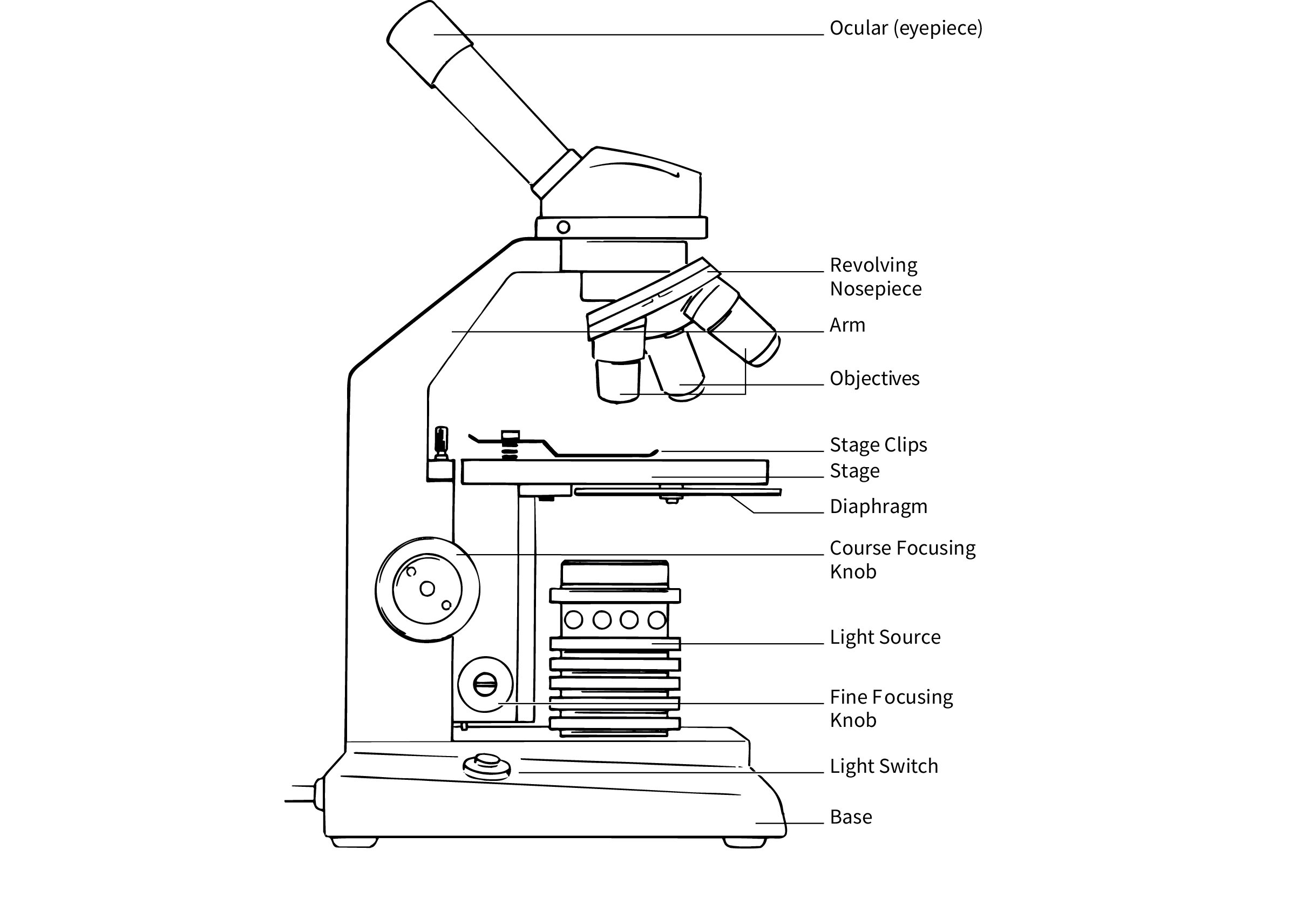
Labelled Diagram of Compound Microscope The below mentioned article provides a labelled diagram of compound microscope. Part # 1. The Stand: The stand is made up of a heavy foot which carries a curved inclinable limb or arm bearing the body tube. The foot is generally horse shoe-shaped structure (Fig. 2) which rests on table top or any other surface on which the microscope in kept.

Dissecting/Stereo microscope | Principle, Parts, working, and ...
Microscope Parts and Functions First, the purpose of a microscope is to magnify a small object or to magnify the fine details of a larger object in order to examine minute specimens that cannot be seen by the naked eye. Here are the important compound microscope parts... Eyepiece: The lens the viewer looks through to see the specimen.

Types of Microscopes: Definition, Working Principle, Diagram ...
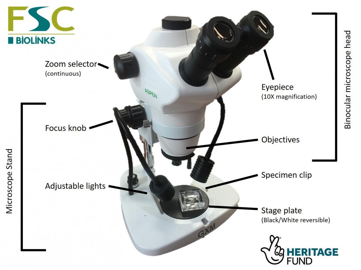
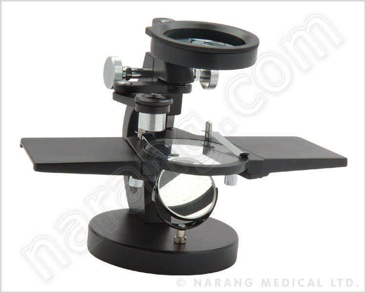
› dissecting-stereoDissecting Stereo Microscope Parts and Functions Dissecting Stereo Microscope Parts and Functions Overview. Also known as a stereoscopic microscope, a dissecting microscope is a type of optical microscope commonly used for studying three-dimensional objects (3-D objects) as well as for dissecting biological specimen (e.g. insects and plant parts etc) at low magnification, between 2 and 100x depending on the microscope.
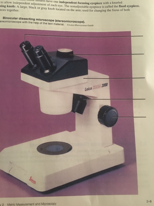
Solved uim e mels have one independent focusing eyepiece ...
Parts of a microscope with functions and labeled diagram - Microbe Notes Figure: Diagram of parts of a microscope There are three structural parts of the microscope i.e. head, base, and arm. Head - This is also known as the body. It carries the optical parts in the upper part of the microscope. Base - It acts as microscopes support. It also carries microscopic illuminators.

Basic Microscopy - An Important Skill for Plant Pathologists
A genetically engineered Plasmodium falciparum parasite vaccine ... WebAug 24, 2022 · Genetically attenuated malaria parasite vaccines are currently being developed as candidates for protection against Plasmodium falciparum infection. Murphy et al. used a genetically attenuated P. falciparum sporozoite vaccine they had previously developed that has deletions in parasite genes P52, P36, and SAP1 (PfGAP3KO) to …

Compound Microscope Parts, Functions, and Labeled Diagram ...
Parts of the Dissecting Microscope - Synonym Dissecting microscopes are used for viewing live specimens or three-dimensional objects too large or thick to be accommodated by compound microscopes. Specimens can be physically manipulated under magnification, since they do not have to be mounted onto a slide for observation under a dissecting microscope. These ...

Parts of a Microscope - SmartSchool Systems
A physical wiring diagram for the human immune system | Nature WebAug 03, 2022 · For imaging, a PerkinElmer Opera Phenix automated spinning-disk confocal microscope was used and each well of a 348-well plate was imaged at 20× magnification with 5 × 5 non-overlapping images ...

Amazon.com : Digital Binocular Stereo Dissecting Microscopes ...
elifesciences.org › articles › 77058A unified view of low complexity regions (LCRs) across species Sep 13, 2022 · The final protein concentration in the reaction was 8.3 μM. After the addition of purified protein, the reaction was mixed by pipetting, 10 μL was loaded onto a microscope slide (Fisher Scientific, 12-544-2), and droplets were immediately imaged using a fluorescent microscope (Evos FL) at 40 X magnification.

Leica Zoom 2000 Stereo Microscope
Microscope, Microscope Parts, Labeled Diagram, and Functions Stage with Stage Clips: The stage of a microscope is a flat platform where you place your subject slides. Stage clips hold the slides in place. The mechanical stage of your microscope will help you to move the slide around by turning two knobs. One knobs moves it left and right, the other knobs moves it up and down.

Dissecting Microscope Uses - New York Microscope Company
› tech-article › uv-properties-ofUV Properties of Plastics: Transmission and Resistance May 03, 2021 · It is at the higher end of energy compared to visible light and is followed in energy by X-rays and the Gamma rays - see diagram. UV radiation is split into three different types as described in table 1 together with their characteristic effect.
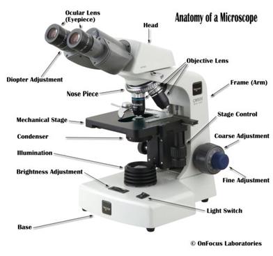
Microscope Parts and Functions
Exemplars tests, practicals & projects - SlideShare WebJun 10, 2013 · Life Sciences/Grade 10 NCS 13 QUESTION 3 3.1 Study the diagram below and answer the questions that follow: 3.1.1 Explain the difference between a food chain and a food web. (2) 3.1.2 In a pyramid of numbers, there is an increase in numbers towards the base of the pyramid. Explain the Biological importance of this concept.

Types of Microscopes: Definition, Working Principle, Diagram ...
What is a Refractometer & How Does it Work - Cole-Parmer WebAug 26, 2022 · Measurements are read at the point where the prism and solution meet. With a low concentration solution, the refractive index of the prism is much greater than that of the sample, creating a large refraction angle and a low reading ("A" on diagram). The reverse would happen with a high concentration solution ("B" on diagram).

20x-40x Industrial Binocular Stereo Microscope PCB Soldering Repairing Tool for Mobile Phone Clock Repairing and PCB Inspection
Single-cell transcriptome profiling reveals neutrophil ... - Nature WebJul 27, 2020 · Specifically, cells would receive corresponding labels with the highest similarity scores, whereas cells with the highest similarity score lower than 0.5 were defined as unassigned.
Dissecting microscope hi-res stock photography and images - Alamy
Dissecting the treatment-naive ecosystem of human melanoma … WebJul 07, 2022 · Introduction. Melanoma brain metastases (MBMs) are the third most common cause of brain metastases after carcinomas of the lung and breast (Eichler et al., 2011) and lead to significant morbidity and mortality (Davies et al., 2011).While treatment with combination immune checkpoint blockade can be effective in patients with MBM (Tawbi …
5 things to consider before purchasing a microscope (for ...
Dissecting microscope (Stereoscopic or Stereo microscope) This microscope is a dual-powered dissecting microscope of 10x-30x with an ability to rotate 360° making it ideal for viewing and focussing better to view samples. By rotating the lenses, users can change the magnification of image.
LAB 1: Scientific Method/Tools of Scientific Inquiry
A Study of the Microscope and its Functions With a Labeled Diagram ... A Study of the Microscope and its Functions With a Labeled Diagram To better understand the structure and function of a microscope, we need to take a look at the labeled microscope diagrams of the compound and electron microscope. These diagrams clearly explain the functioning of the microscopes along with their respective parts.
Dissecting Microscopes | Senior Dissecting Microscopes ...

Dissecting Microscope Parts And Functions. All You Need To Know

Microscope Parts & Functions - AmScope

How To Draw A Microscope, Step by Step, Drawing Guide, by ...
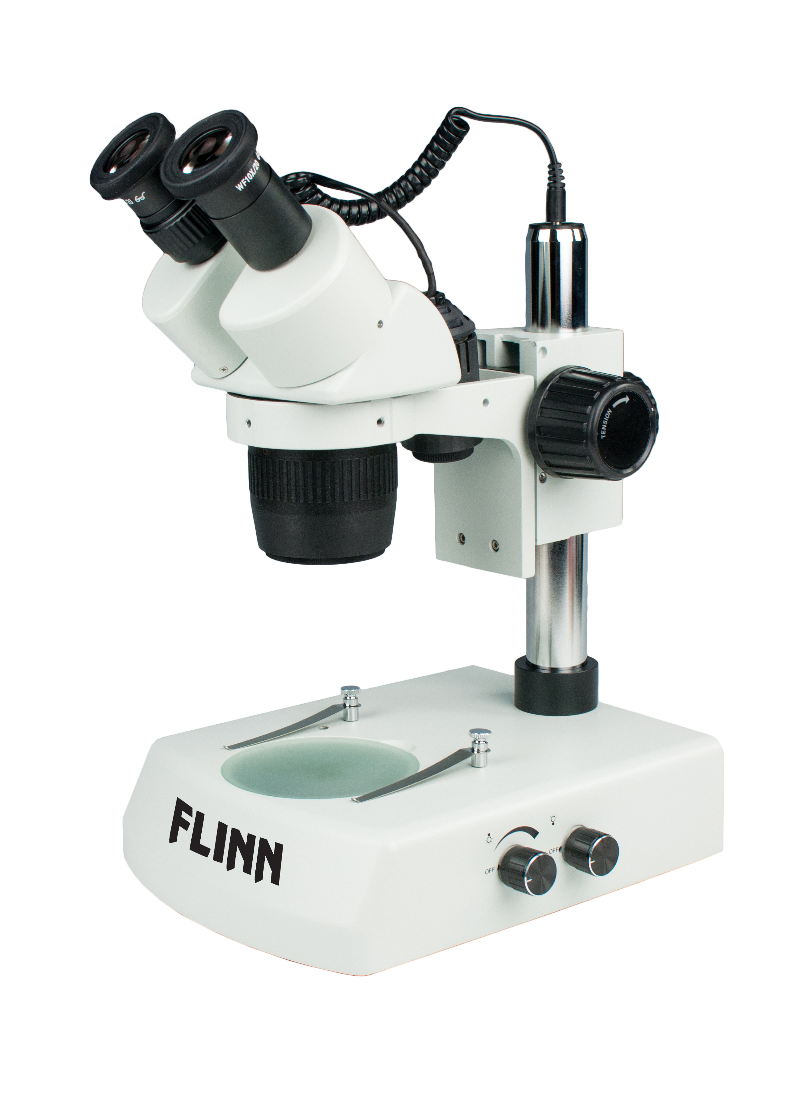
What is a Compound Microscope? | Flinn Scientific
Stereoscopes and Dissecting Microscopes for Biology and Life ...

Microscopes. (a) Binocular dissecting microscope. (b ...
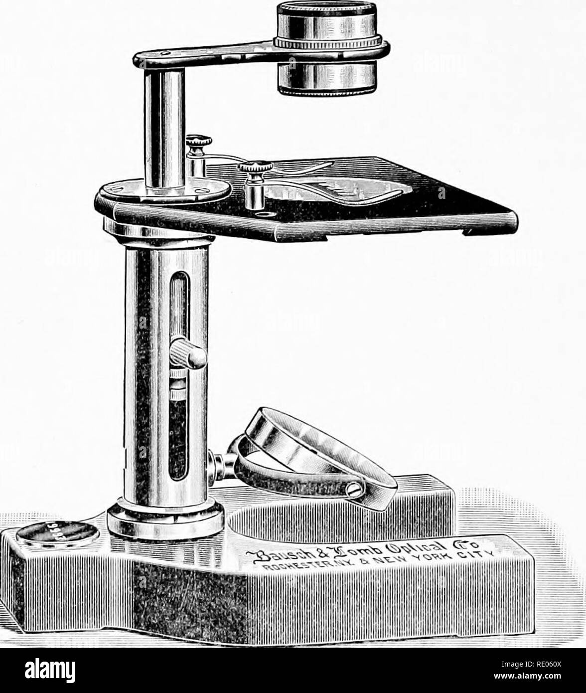
The microscope; an introduction to microscopic methods and to ...
Dissecting Stereo Microscope Parts and Functions
Parts of Stereo Microscope (Dissecting microscope) – labeled ...

Microscope With Labels clip art | Microscope parts, Science ...

Compound and Stereo- microscopes - Microscopes 4 Schools
Dissecting Microscopes - ppt download

Microscope World Blog: Dissecting Microscopes
How to Use a Stereo Microscope and Science Lesson Ideas
PRACTICAL BOOKLET - BIOLOGY4ISC

Swift® SM100 Series Use and Care Manual | Carolina.com
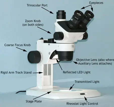
Stereo Microscope - Parts, Types and Uses - Laboratoryinfo.com
Stereo Microscope: Uses, Advantages, and Disadvantages ...

Parts of Stereo Microscope (Dissecting microscope) – labeled ...

Microscope | Dissecting microscope, Microscope, Microscope parts

Microscope World Blog: November 2013

Tsetse biology, systematics and distribution, techniques

Explain Dissecting microscope - Brainly.in

How to Draw a Simple Microscope Diagram
Post a Comment for "45 dissecting microscope diagram with labels"