38 cell membrane diagram with labels
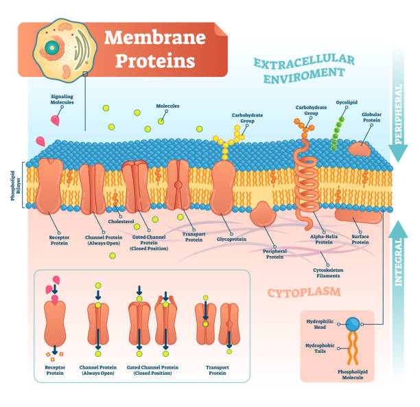
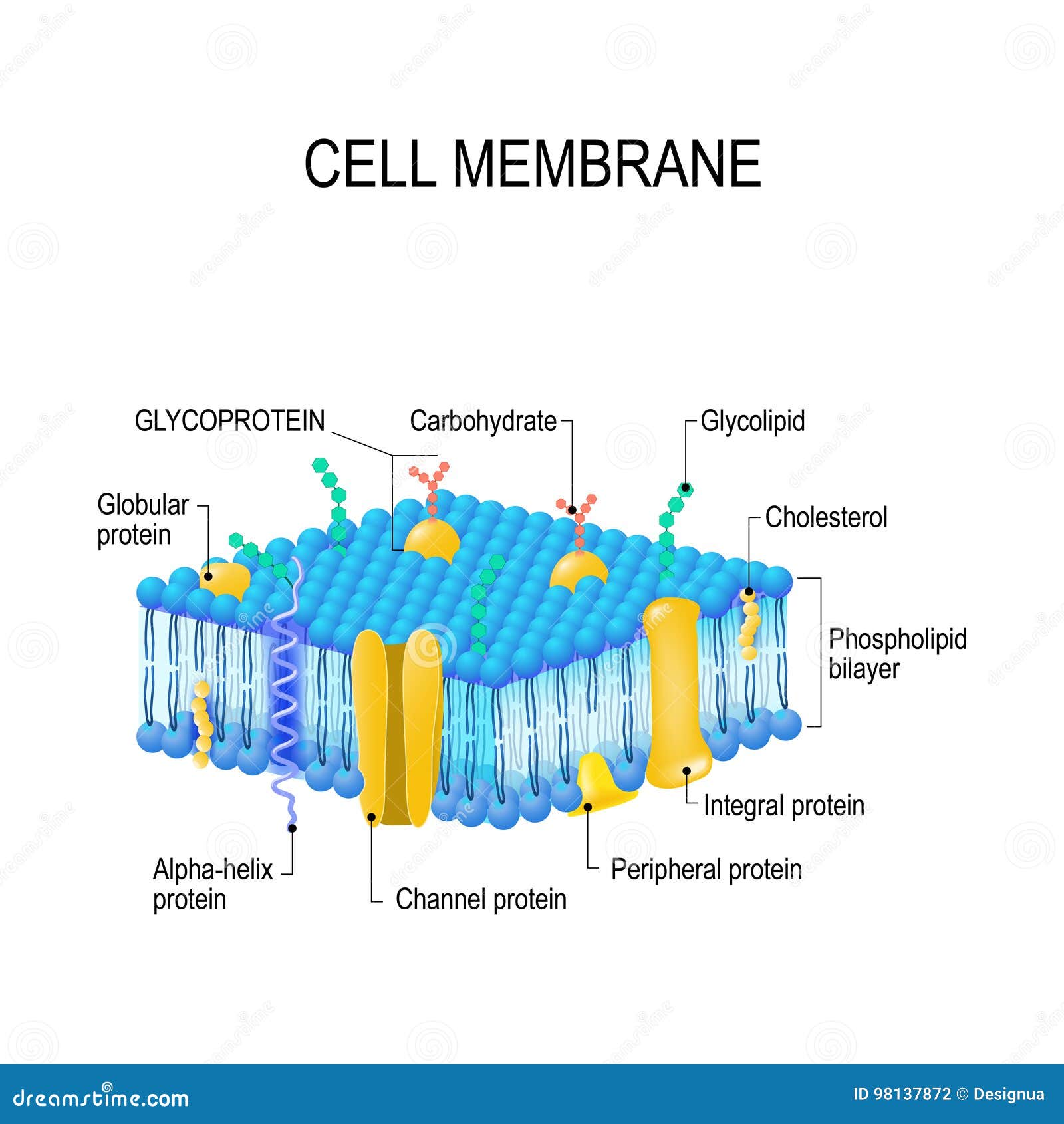
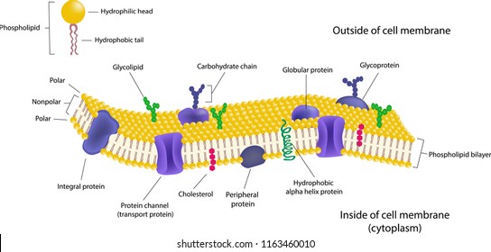
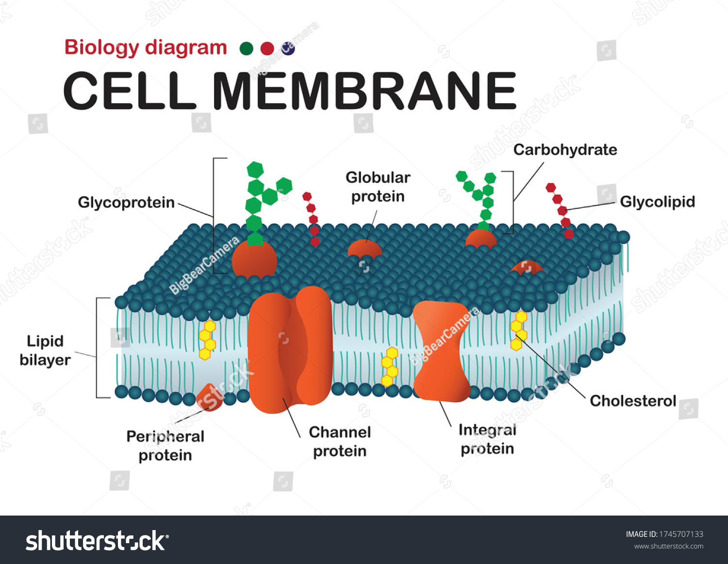
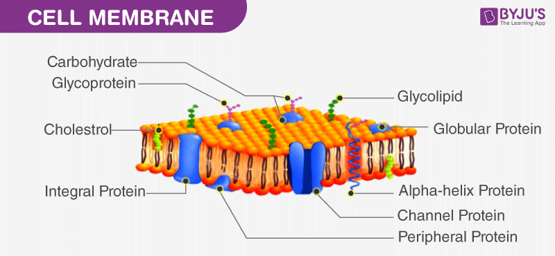
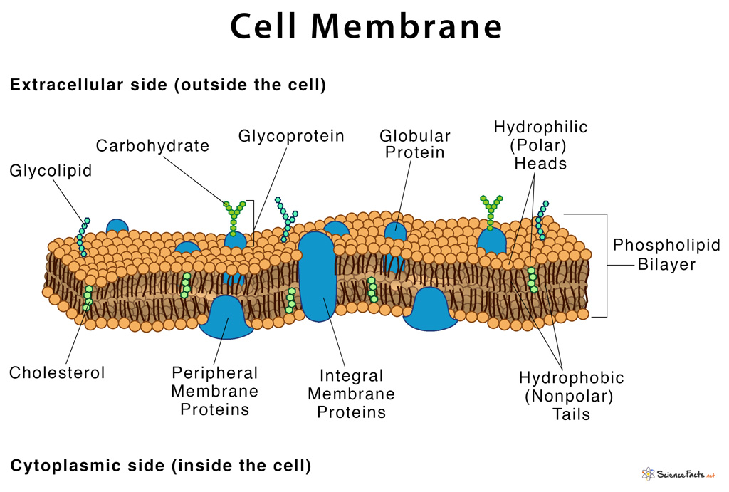
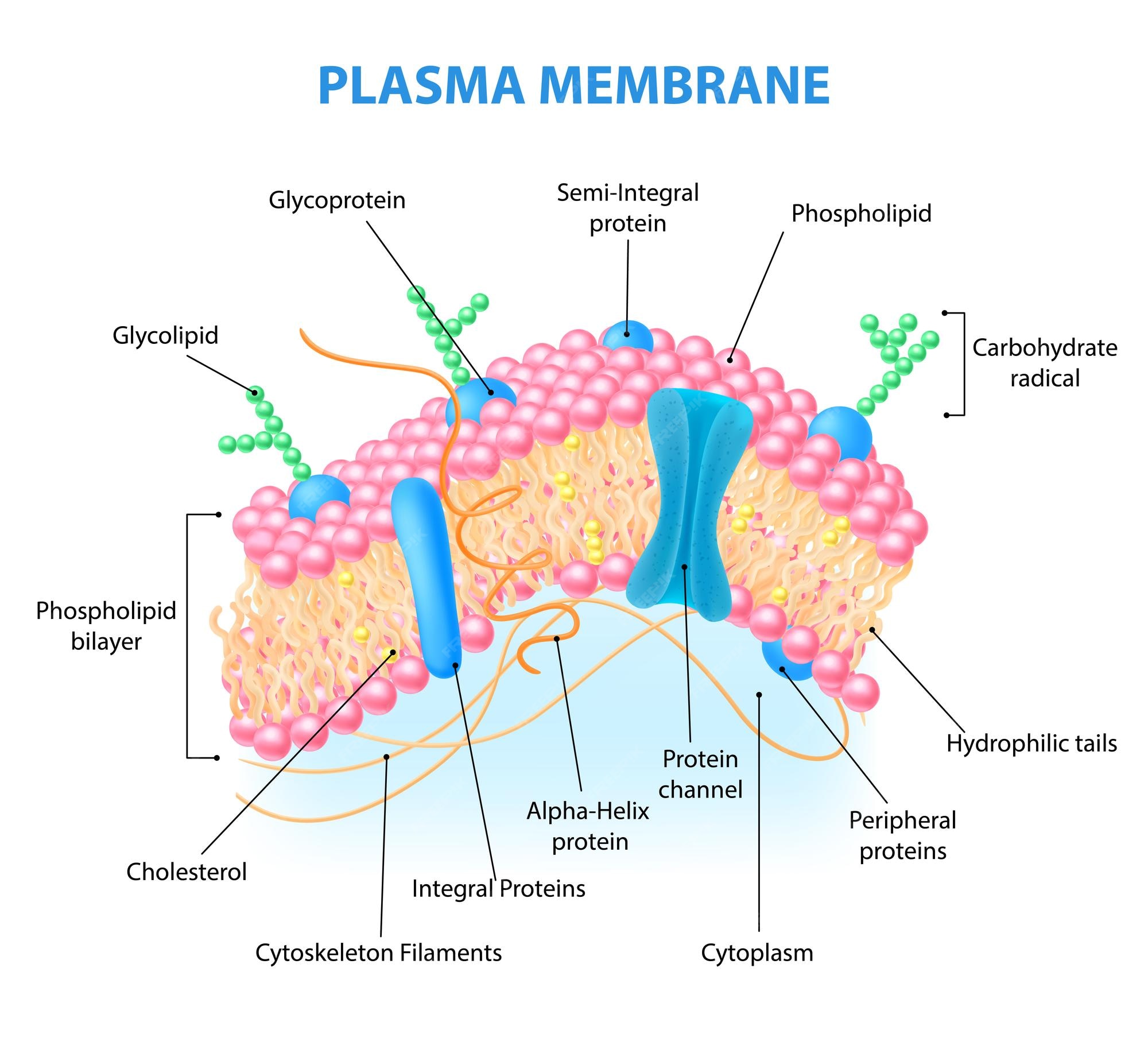
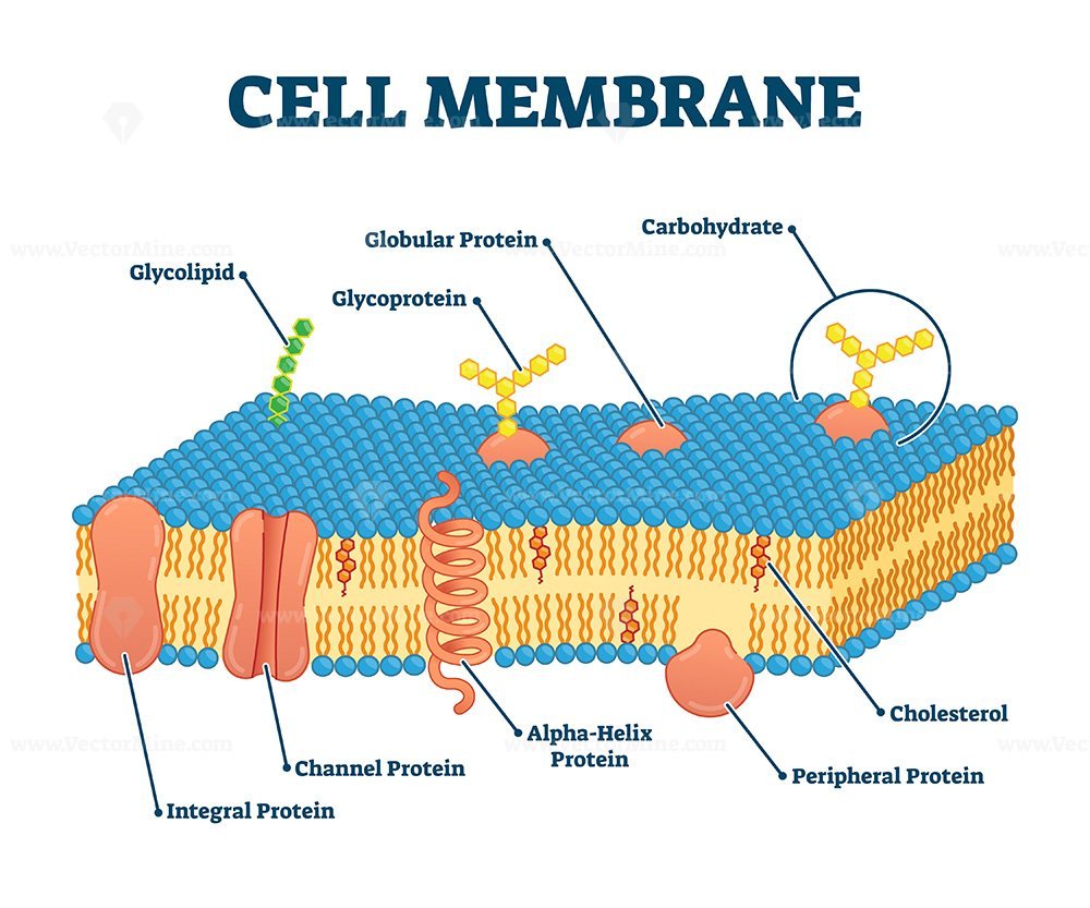
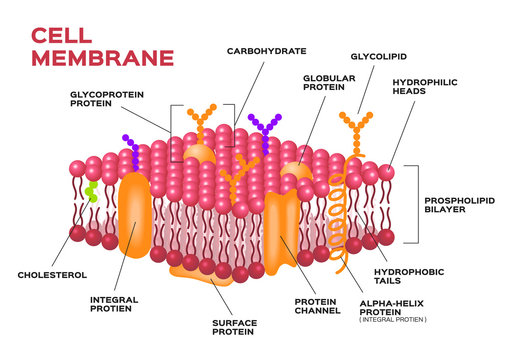
Structure of the plasma membrane (article) | Khan Academy The principal components of the plasma membrane are lipids (phospholipids and cholesterol), proteins, and carbohydrate groups that are attached to some of the lipids and proteins. A phospholipid is a lipid made of glycerol, two fatty acid tails, and a phosphate-linked head group. Cell Membrane Labeled | EdrawMax Template Mar 2, 2022 ... In the cell membrane labeled diagram, we see Glycolipid, Glycoprotein, Globular Protein, Carbohydrate, Cholesterol, Peripheral Protein, ...
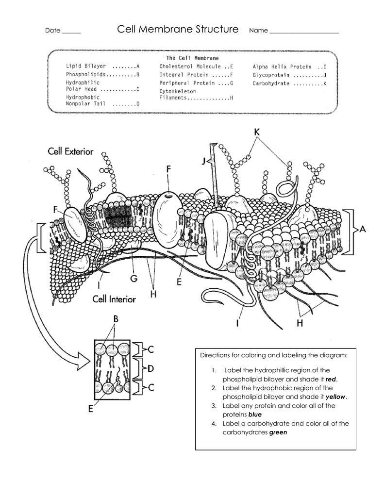
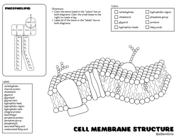
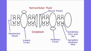
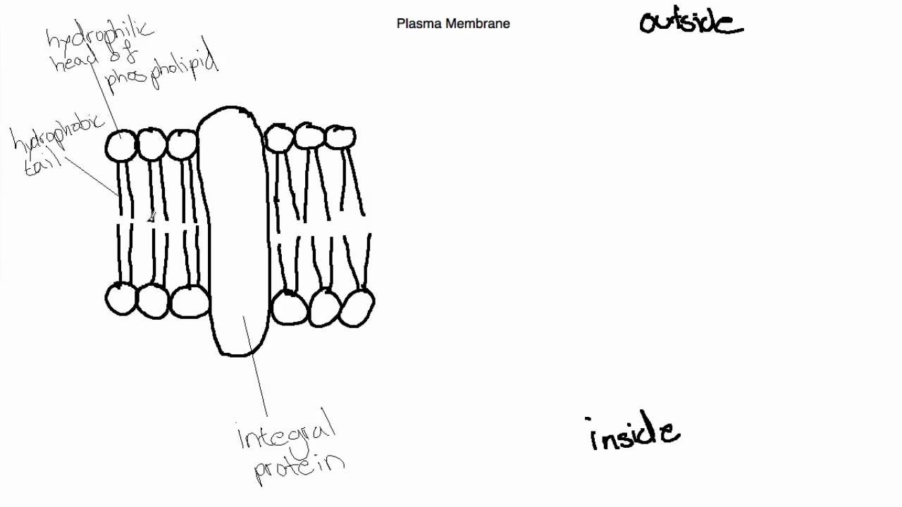
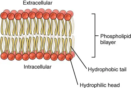
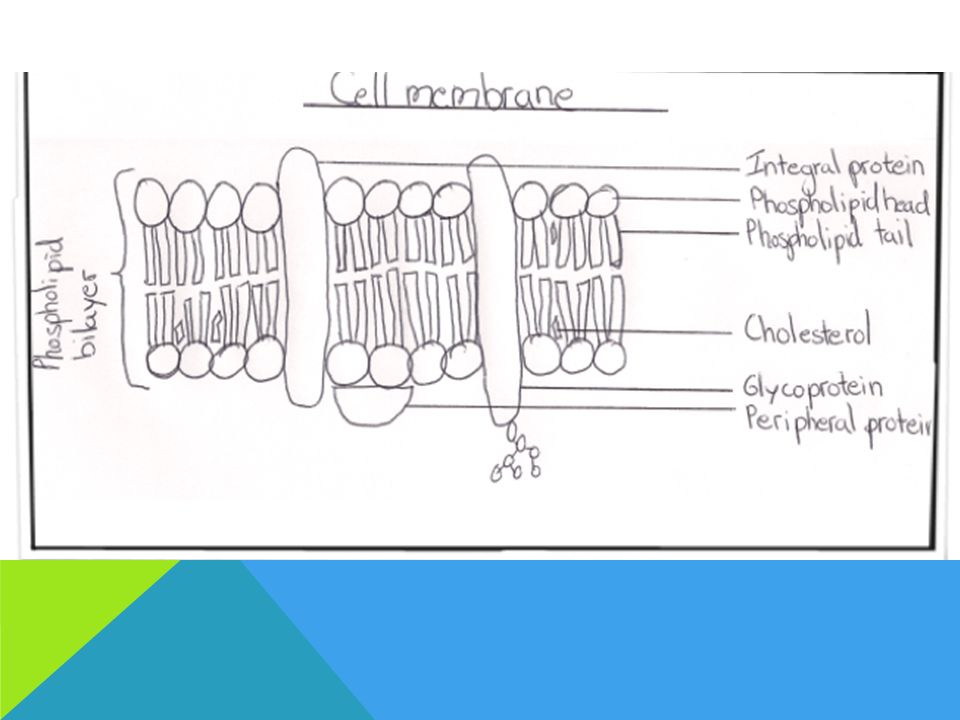
2.4.1 Draw and label a diagram to show the structure of membranes Apr 25, 2012 ... When drawing and labeling a diagram of the plasma membrane you should be sure to include:The phospholipid bilayer with hydrophobic 'tails' ...

Cell membrane diagram with labels
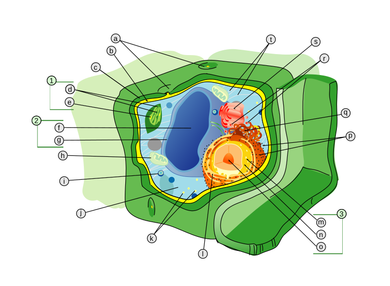
PDF Human Cell Diagram, Parts, Pictures, Structure and Functions The cell membraneis the outer coating of the cell and contains the cytoplasm, substances within it and the organelle. It is a double-layered membrane composed of proteins and lipids. The lipid molecules on the outer and inner part (lipid bilayer) allow it to selectively transport substances in and out of the cell. Endoplasmic Reticulum Cell Membrane Function and Structure - ThoughtCo The cell membrane (plasma membrane) is a thin semi-permeable membrane that surrounds the cytoplasm of a cell. Its function is to protect the integrity of the interior of the cell by allowing certain substances into the cell while keeping other substances out. Human Cell Diagram, Parts, Pictures, Structure and Functions Diagram of the human cell illustrating the different parts of the cell. Cell Membrane. The cell membrane is the outer coating of the cell and contains the cytoplasm, substances within it and the organelle. It is a double-layered membrane composed of proteins and lipids. The lipid molecules on the outer and inner part (lipid bilayer) allow it to ...
Cell membrane diagram with labels. Labeled Diagram Of Cell Membrane : Electron Micrograph The nucleus and mitochondria are two examples. Copy of labeling cell membrane labelled diagram. Some of the major parts of the plasma membrane are : Phospholipid bilayer · phospholipid bilayer ; It supports and helps maintain a cell's shape. 1)cell membrane 2)vacuole 3)nucleus 4)endoplasmic reticulum 5)mitochondria 6)golgi body. › articles › s41586/021/03941-1Morphological diversity of single neurons in molecularly ... Oct 06, 2021 · Dendritic and axonal morphology reflects the input and output of neurons and is a defining feature of neuronal types1,2, yet our knowledge of its diversity remains limited. Here, to systematically ... Schematic Diagram of a Cell Membrane - Pinterest Schematic Diagram of a Cell Membrane Cell Membrane Structure, Plasma Membrane, Extracellular Fluid, ... Membrane proteins labeled vector illustration. Diagram of a cell membrane with labels - nist.gov Essential Biological FunctionsImmune response, Cell metabolism, Neurotransmission, Photosynthesis, Cell adherence, Cell growth and differentiationPotential Commercial ApplicationsDrug response monitoring, Chemical manufacturing, Biosensing, Energy conversion, Tissue engineering ... Diagram of a cell membrane with labels. Appears In. Biology in ...
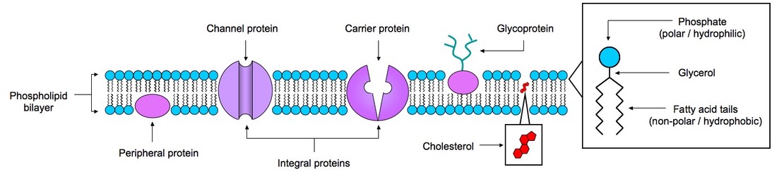
Basic Cell Membrane Label - Labelled diagram - Wordwall Integral Protein (channel), Peripheral Protein, Phosphate, Lipid, Hydrophilic, Hydrophobic, Glycoprotein. Cell Membrane Labeling | Cell Structure Quiz - Quizizz Question 9. 30 seconds. Q. The function of carbohydrates in the cell membrane is to. answer choices. stick to other cells and sense stuff outside the cell. create energy for the cell. form the phospholipid bilayer. allow molecules to pass through it. Cell membrane with labeled educational structure scheme vector ... 2. Editable Vector .EPS-10 file. 3. High-resolution JPG image. Use for everything except reselling item itself. Description: Cell membrane with labeled educational structure scheme vector illustration. Anatomical closeup drawing with cross section element. Carbohydrate, globular protein or cholesterol location visualization. Cell Membrane - The Definitive Guide | Biology Dictionary The cell membrane, also known as the plasma membrane, is a double layer of lipids and proteins that surrounds a cell. It separates the cytoplasm (the contents of the cell) from the external environment. It is a feature of all cells, both prokaryotic and eukaryotic. a 3D diagram of the cell membrane Function of the Cell Membrane
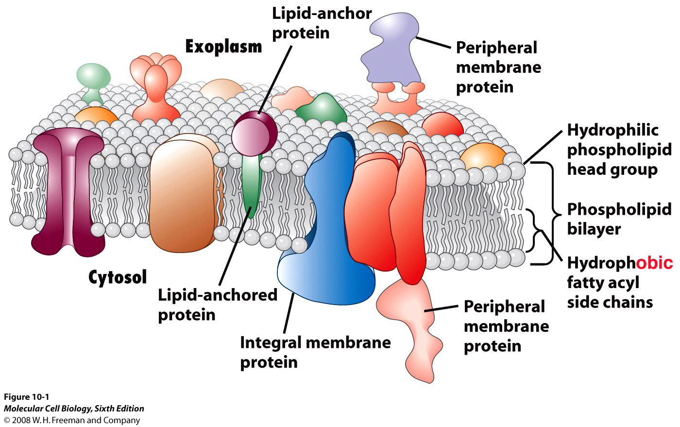
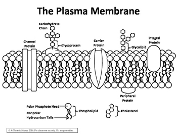
PDF Membrane Structure and Function - Phoenix College the cell membrane. Major Components of the Cell Membrane: Lipids • Phospholipids are amphipathic molecules (with hydrophobic tails and a hydrophilic head) • One of the phospholipid tails exist mostly in a trans configuration, providing more fluidity to the membrane • Cholesterol is a rigid Draw and label a simple line diagram of a cell membrane. Include ... The outer covering of the body cells, which maintains homeostatic condition between inside and outside of the cell is called cell membrane. It is made up of ... File : Cell membrane detailed diagram id.svg - Wikimedia Original upload log []. This image is a derivative work of the following images: File:Cell_membrane_detailed_diagram_en.svg licensed with PD-user . 2009-02-23T18:08:26Z Bibi Saint-Pol 877x361 (487132 Bytes) {{Information |Description= {{en|The cell membrane, also called the plasma membrane or plasmalemma, is a semipermeable lipid bilayer common to all living cells. Structure of Membrane in Cells (With Diagram) - Biology Discussion In Gram-positive bacteria, the cell wall (Fig. 2.11) is (30-100 nm) thick outside the plasma membrane. The cell wall consists of peptidoglycan which is a polysaccharide- peptide complex. The polysaccharide complex of the adjacent chains are joined together by peptide bridges containing different kinds of amino acids like D and L-alanine, D ...
Animal Cell- Definition, Structure, Parts, Functions, Labeled ...
Cell Organelles- Definition, Structure, Functions, Diagram - Microbe Notes A cell wall is multilayered with a middle lamina, a primary cell wall, and a secondary cell wall. The middle lamina contains polysaccharides that provide adhesion and allow binding of the cells to one another. After the middle lamina is the primary cell wall which is composed of cellulose.
Cell Membrane Structure
Label the Cell Membrane - Labelled diagram - Wordwall Label the Cell Membrane - Labelled diagram. Home. Features. Contact. Price Plans. Log In. Sign Up. Language. channel protein, cholesterol, external cell environment, hydrophilic (water loving) part of phospholipid bilayer, peripheral protein, internal environment of the cell, hydrophobic (water fearing) part of phospholipid bilayer, glycolipid.
2,000 Human Cell Membrane Stock Photos, Pictures & Royalty ...
Label Cell Membrane Diagram | Quizlet Label Cell Membrane Diagram | Quizlet Label Cell Membrane 3.5 (22 reviews) + − Learn Test Match Created by Shinaala20 Terms in this set (8) Integral Protein ... Phospholipid Bilayer ... Glycoprotein ... Phosphate Heads ... Cholesterol ... Fatty Acid Tails ... Glycolipids ... Peripheral Protein ... lara2151 8 terms
Cell Membrane Stock Illustrations – 5,888 Cell Membrane Stock ...
quizlet.com › 525123594 › week-3-membrane-transportWeek 3: Membrane Transport Flashcards | Quizlet The cellular plasma membrane is selectively permeable, which means some materials move through it while others cannot. The movement of materials into and out of the cell is called membrane transport. This activity will help you identify the different mechanisms of membrane transport.
Cell membrane - Teaching resources
Solved Match the cell membrane structure or its function - Chegg Transcribed image text: Match the cell membrane structure or its function with the correct letter from the diagram Label the hydrophobic and hydrophobic portions of the phospholipids. Attracts water Phospholipid bilayer Repels water Helps maintain flexibility of membrane Makes up the bilayer (2 answers) Involved in cell-to-cell recognition WHITE Helps transport materials (such as glucose ...
Cell/Plasma Membrane Structure Color & Label Perfect for Interactive Notebooks!
CELL MEMBRANE LABEL Diagram | Quizlet Practice labeling the parts of the cell membrane Terms in this set (6) Channel Protein hole or tunnel that particles may pass through to go in / out of cell Marker protein identifies or labels the cell Receptor protein receives information Heads part of the phospholipid that loves water (hydrophili) - points to the most outside and inside of cell
14,993 Cell Membrane Images, Stock Photos & Vectors ...
achieverpapers.comAchiever Papers - We help students improve their academic ... 100% money-back guarantee. With our money back guarantee, our customers have the right to request and get a refund at any stage of their order in case something goes wrong.

ImageQuiz: Cell Membrane Labeling
› cells › bactcellInteractive Bacteria Cell Model - CELLS alive They have an outer cell wall that gives them shape. Just under the rigid cell wall is the more fluid cell membrane. The cytoplasm enclosed within the cell membrane does not exhibit much structure when viewed by electron microscopy. Use the following animation to explore bacterial structure.
2.4.1 Draw and label a diagram to show the structure of ...
Schematic Diagram of a Cell Membrane - Pinterest Honors Biology @ Lawrenceville: cells Cell Membrane Structure, Plasma Membrane, Extracellular Fluid, ... Membrane proteins labeled vector illustration.
Membranes"
Cell Membrane (Plasma Membrane) - Genome.gov The cell membrane, also called the plasma membrane, is found in all cells and separates the interior of the cell from the outside environment. The cell membrane consists of a lipid bilayer that is semipermeable. The cell membrane regulates the transport of materials entering and exiting the cell. Narration 00:00 …
2.4 Membranes | BioNinja
Cell: Structure and Functions (With Diagram) - Biology Discussion Eukaryotic Cells: 1. Eukaryotes are sophisticated cells with a well defined nucleus and cell organelles. 2. The cells are comparatively larger in size (10-100 μm). 3. Unicellular to multicellular in nature and evolved ~1 billion years ago. 4. The cell membrane is semipermeable and flexible. 5. These cells reproduce both asexually and sexually.

a. Draw and label a simple line diagram of a cell membrane ...
A Labeled Diagram of the Animal Cell and its Organelles A Labeled Diagram of the Animal Cell and its Organelles There are two types of cells - Prokaryotic and Eucaryotic. Eukaryotic cells are larger, more complex, and have evolved more recently than prokaryotes. Where, prokaryotes are just bacteria and archaea, eukaryotes are literally everything else.
Biology Diagram Show Structure Cell Membrane Stock Vector ...
assignmentessays.comAssignment Essays - Best Custom Writing Services Get 24⁄7 customer support help when you place a homework help service order with us. We will guide you on how to place your essay help, proofreading and editing your draft – fixing the grammar, spelling, or formatting of your paper easily and cheaply.

The Plasma Membrane | Plasma membrane, Biology classroom ...
Animal Cell Diagram: Functions & Structure - Collegedunia Animal cells are eukaryotic cells that are seen specifically in animal tissues. It is characterised by the absence of cell wall, with cell organelles enclosed within the membrane of the cell .They contain membrane-bound nuclei. The diagram of animal cell is beneficial in understanding the structure and functions of an animal.
IB Biology Topic 2.4.1 Draw and Label the Plasma Membrane
Simple Columnar Epithelium: A Labeled Diagram and Functions These form a brush border. They also increase the absorptive surface area of these cells. On a concluding note, simple columnar epithelium has two primary functions of absorption and secretion. In the small intestine, it facilitates the absorption of nutrients. It also secretes mucus, which helps to lubricate, moisten, and protect the surface.
Cell Membrane Explained: Here's Everything You Need to Know ...
Cell Diagrams with Labelling Activity - Learnful The cell structure illustrations for these diagrams were generated in BioRender. Both diagrams feature a drag-and-drop labelling activity created with H5P here ...

Cell Membrane Structure and Function - Biology Wise
en.wikipedia.org › wiki › Cell_nucleusCell nucleus - Wikipedia The cell nucleus (pl. nuclei; from Latin nucleus or nuculeus, meaning kernel or seed) is a membrane-bound organelle found in eukaryotic cells.Eukaryotic cells usually have a single nucleus, but a few cell types, such as mammalian red blood cells, have no nuclei, and a few others including osteoclasts have many.
Cell Wall and Cell Membrane- Structure, Functions and Differences
Plasma Membrane Function, Structure & Diagram - Study.com 2. property of the plasma membrane that allows some substances into the cell and keeps others out 4. main structural component of the plasma membrane 6. nonpolar part of a phospholipid 11. protein...

Cell Membrane (Labeling, pt. 1) - Biology Honors - Champagne ...
Plant Cell: Diagram, Types and Functions - Embibe Exams Xylem is a tissue that is formed of four different types of cells, i.e. tracheids, xylem vessels, xylem fibres and xylem parenchyma. They are the transport cells in vascular plants. They help in the transport of water and minerals from the roots to the leaves and other parts of the plants. The movement of water is unidirectional. Phloem

Plasma membrane worksheet
Labeled Plant Cell With Diagrams | Science Trends The parts of a plant cell include the cell wall, the cell membrane, the cytoskeleton or cytoplasm, the nucleus, the Golgi body, the mitochondria, the peroxisome's, the vacuoles, ribosomes, and the endoplasmic reticulum. Parts Of A Plant Cell The Cell Wall Let's start from the outside and work our way inwards.
Cell wall - Wikipedia
Animal Cells: Labelled Diagram, Definitions, and Structure - Research Tweet The endoplasmic reticulum (s) are organelles that create a network of membranes that transport substances around the cell. They have phospholipid bilayers. There are two types of ER: the rough ER, and the smooth ER. The rough endoplasmic reticulum is rough because it has ribosomes (which is explained below) attached to it.

Cell Membrane Model - Perkins School for the Blind
Labeling cell membrane - Teaching resources - Wordwall 3048 results for 'labeling cell membrane'. Labeling Cell Membrane Labelled diagram. by Walukas.

Label Cell Membrane Diagram | Quizlet
en.wikipedia.org › wiki › History_of_cell_membraneHistory of cell membrane theory - Wikipedia Two experiments in 1924 laid the groundwork to fill in this gap. By measuring the capacitance of erythrocyte solutions Fricke determined that the cell membrane was 3.3 nm thick. Although the results of this experiment were accurate, Fricke misinterpreted the data to mean that the cell membrane is a single molecular layer.

Learn About Diagram Of Plasma Membrane | Chegg.com
Cell Membrane Structure and Function - Biology Wise Cell membrane is a protective covering that acts as a barrier between the inner and outer environment of a cell (in animals). In plant cells, the membrane encapsulates the protoplasm. This organelle is also referred to as plasma membrane. Images obtained through electron micrography reveal the bilayer structure of cell membranes.
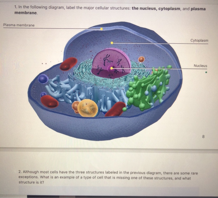
Solved 1. In the following diagram, label the major cellular ...
Human Cell Diagram, Parts, Pictures, Structure and Functions Diagram of the human cell illustrating the different parts of the cell. Cell Membrane. The cell membrane is the outer coating of the cell and contains the cytoplasm, substances within it and the organelle. It is a double-layered membrane composed of proteins and lipids. The lipid molecules on the outer and inner part (lipid bilayer) allow it to ...
Cell Membrane: Definition, Structure, & Functions with Diagram
Cell Membrane Function and Structure - ThoughtCo The cell membrane (plasma membrane) is a thin semi-permeable membrane that surrounds the cytoplasm of a cell. Its function is to protect the integrity of the interior of the cell by allowing certain substances into the cell while keeping other substances out.

Draw and label a simple line diagram of a cell membrane ...
PDF Human Cell Diagram, Parts, Pictures, Structure and Functions The cell membraneis the outer coating of the cell and contains the cytoplasm, substances within it and the organelle. It is a double-layered membrane composed of proteins and lipids. The lipid molecules on the outer and inner part (lipid bilayer) allow it to selectively transport substances in and out of the cell. Endoplasmic Reticulum
BIOLOGY 11 IB 2.4: MEMBRANES. ASSESSMENT STATEMENTS 2.4.1Draw ...
Schematic illustration of N 3 -labeled T cell membrane ...
Premium Vector | Realistic human cell anatomy infographics ...
Cell membrane with labeled educational structure scheme vector illustration
Plasma Membrane / Fluid Mosaic Diagram
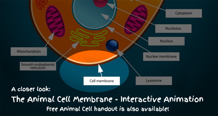
Animal Cell Membrane - Interactive DiagramkidCourses.com

File:Cell membrane detailed diagram en.svg - Wikimedia Commons

Fluid Mosaic Model Vector Illustration. Labeled Cell Membrane ...
Cell Membrane Images – Browse 10,623 Stock Photos, Vectors ...

Membranes

1. Labeling a cell membrane Diagram | Quizlet
Post a Comment for "38 cell membrane diagram with labels"